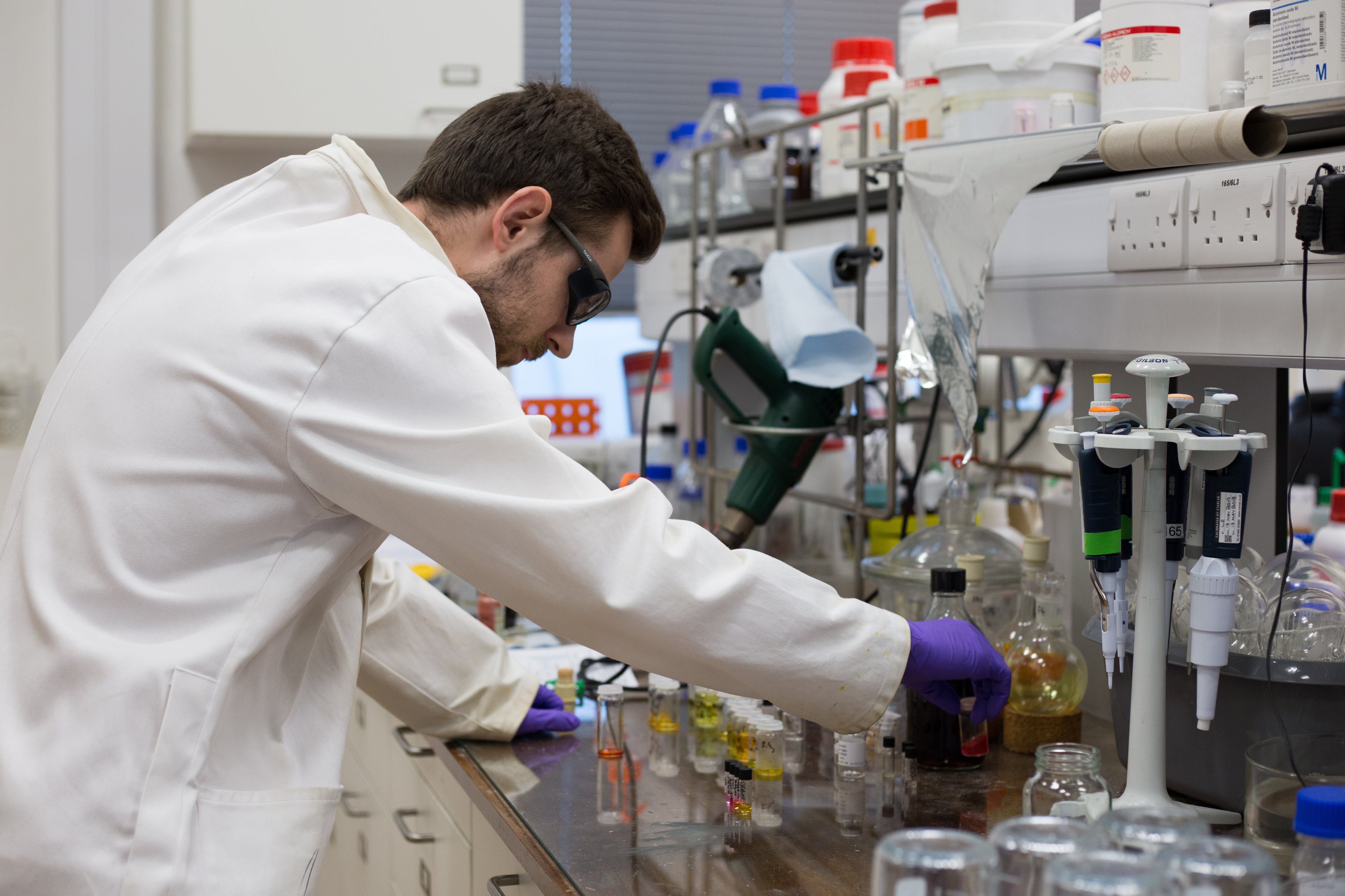
Our researchers have a range of equipment and tools available to them across different departments at the University. A database of major tools and facilities is found on http://www.equipment.admin.cam.ac.uk/
Some important research tools are made available to us by the Cambridge facility of the Henry Royce Institute, based out of the Maxwell Centre – a list of these is included below.
Further information including booking details can be found on the Maxwell Centre website.
Features:
- Standard (Ac/Contact Mode)
- Piezo Force Microscopy
- Magneto Force Microscopy
- Fluid Cell (closed fluid exchanges whilst imaging)
- Electrical Measurements
- Electrostatic Force Microscopy
- Scanning Kelvin Probe
- Conductive AFM (ORCA)
Location: Maxwell Lab 0.83
Contacts: Eduardo Camarillo Abad (ec675) / Anna Scheeder (afs41)
Features
- Size range maximum diameter 0.3nm to 10 microns
- Molecular weight range 980Da to 2x1E7Da
- Zeta potential range > +/- 500mV
- Mobility range >+/- 20µ.cm/V.s
Location: Maxwell Lab 0.80
Contact: Anna Scheeder (afs41) / Reece McCoy (rm991)
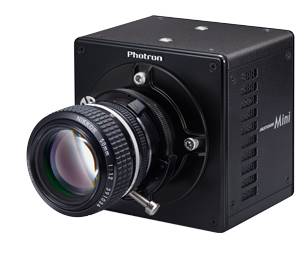
Features:
- Triggers before, during or after capture
- Microscope adapter
- Versatile clamp arm
- Strong light
- Macro lens
- Photo-processing software
Location: Maxwell 0.80
Contact: Oliver Powell (ofjp2)
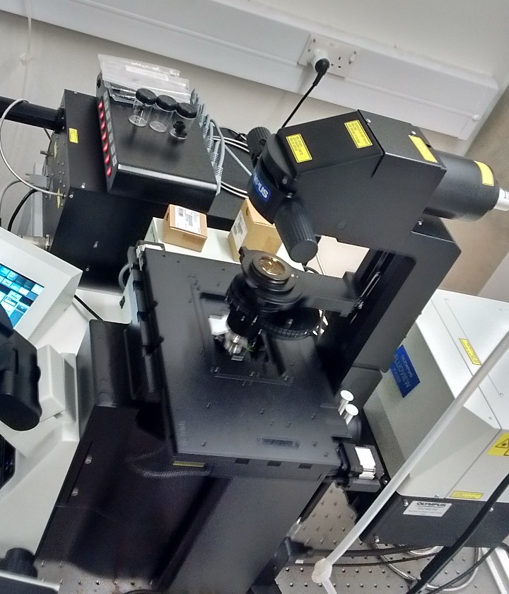
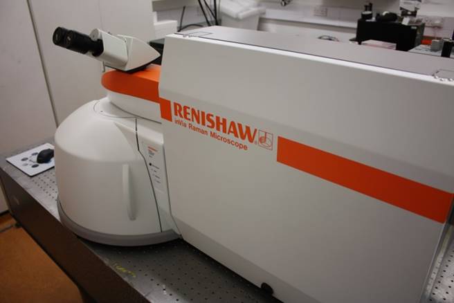
Features:
- Excitation sources: 532nm, 633nm (interchangeable)
- Integrated research grade microscope
- Highly automated
- High resolution confocal map scans
- Depth scans
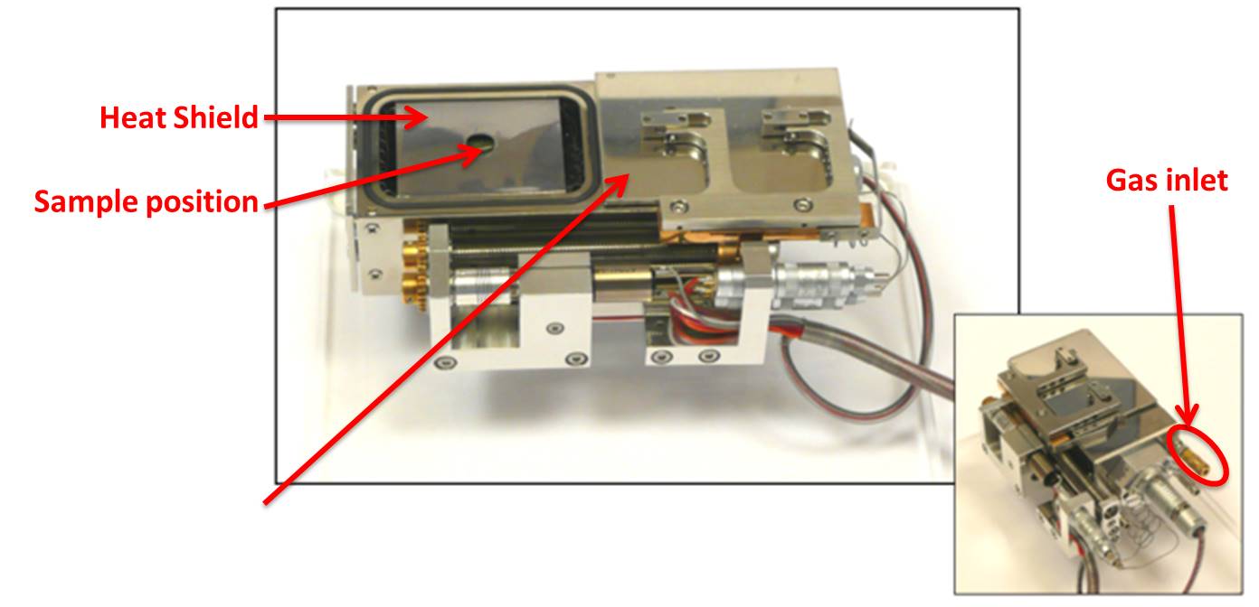
Features:
- Use with ZEISS Gemini 300 Environmental Scanning Electron Microscope
- Temperatures from RT to 1050°C
- Local gas dosing through inlet
- Suitable for use in humid, and corrosive environments
Location: CAPE Room 49 (Ground floor)
Contact: Jinfeng Yang (jy441)
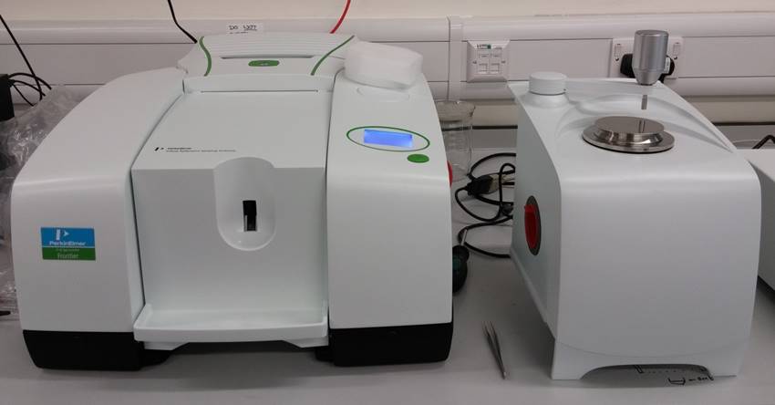
IR spectroscopy of chemicals and nanomaterials (analysis of functional groups on nanoparticles, contaminants, evolution of chemical reactions)
Location: IfM AR-1-002
Contact: Alasdair Tew (at929)
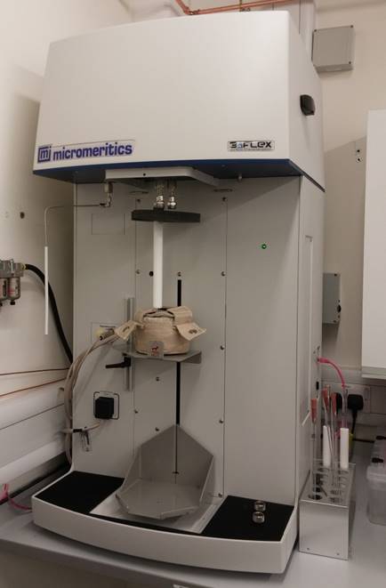
High-performance physisorption – analysis of surface area, pore size distribution of nanoparticles, MOFS, zeolites, etc.
Location: IfM ARG-011
Contact: Alasdair Tew (at929)
Features:
- High resolution, fluorescence imaging, FRAP, FRET, time-lapse, 3D
- imaging, mosaic and multi-point imaging
Location: Maxwell Lab 0.81
Contact: Roger Rubio Sanchez (rmr44); Ashleigh Ruane (ar2060); Andrei Paraschiv (aap69)
Features:
- Standard (Ac/Contact Mode)
- Piezo Force Microscopy
- Magneto Force Microscopy
- Fluid Cell (closed fluid exchanges whilst imaging)
- Electrical Measurements
- Electrostatic Force Microscopy
- Scanning Kelvin Probe
- Conductive AFM (ORCA)
Location: Maxwell Lab 0.83
Contacts: Eduardo Camarillo Abad (ec675) / Anna Scheeder (afs41)
Features
- Size range maximum diameter 0.3nm to 10 microns
- Molecular weight range 980Da to 2x1E7Da
- Zeta potential range > +/- 500mV
- Mobility range >+/- 20µ.cm/V.s
Location: Maxwell Lab 0.80
Contact: Anna Scheeder (afs41) / Reece McCoy (rm991)
Features:
- Triggers before, during or after capture
- Microscope adapter
- Versatile clamp arm
- Strong light
- Macro lens
- Photo-processing software
Location: Maxwell 0.80
Contact: Oliver Powell (ofjp2)
Features:
- Excitation sources: 532nm, 633nm (interchangeable)
- Integrated research grade microscope
- Highly automated
- High resolution confocal map scans
- Depth scans
Features:
- Use with ZEISS Gemini 300 Environmental Scanning Electron Microscope
- Temperatures from RT to 1050°C
- Local gas dosing through inlet
- Suitable for use in humid, and corrosive environments
Location: CAPE Room 49 (Ground floor)
Contact: Jinfeng Yang (jy441)
IR spectroscopy of chemicals and nanomaterials (analysis of functional groups on nanoparticles, contaminants, evolution of chemical reactions)
Location: IfM AR-1-002
Contact: Alasdair Tew (at929)
High-performance physisorption – analysis of surface area, pore size distribution of nanoparticles, MOFS, zeolites, etc.
Location: IfM ARG-011
Contact: Alasdair Tew (at929)
Features:
- High resolution, fluorescence imaging, FRAP, FRET, time-lapse, 3D
- imaging, mosaic and multi-point imaging
Location: Maxwell Lab 0.81
Contact: Roger Rubio Sanchez (rmr44); Ashleigh Ruane (ar2060); Andrei Paraschiv (aap69)
For admissions related queries, please read the information on our admissions and course details pages before contacting us on nanodtc.admissions@nanodtc.cam.ac.uk.
Academics and Prospective Industry Partners should contact our Deputy Director, Dr. Karishma Jain.
For enquiries related to equipment access, please read the details on our equipment page before contacting the relevant person.
For outreach related queries, please contact external@nanodtc.cam.ac.uk
For all other enquiries, please contact the NanoDTC Administrator by email.
Further research tools are available across different departments. Some of these are available as shared research facilities, while others require approval of the relevant group leader.
Department of Physics
SEM, TEM, FIB, ESEM, Ultra Microtome
Optoelectronics, Nanophotonics and Microelectronics groups
Various device fabrication and Optical measurement tools
Optical tweezers, Image analysis, Emulsion PCR, Confocal Microscopy, Microfluidics
Scanning Kerr Microscopy, SQUID, Electroluminescence, Quantum conductance
Clean rooms, Molecular Beam Epitaxy, Electron Beam Lithography
Department of Chemistry
TGA, DSC, GPC, AFM, Spectrascopic ellipsometry, Viscometry FT-IR, Cyclic voltammetry
Optical/Microcantilever/CNT Sensors, Raman and fluorescence spectroscopy, Microfluidics
NMR, BET, High Temp Ovens, Impedance Spectroscopy, Glove Box, Ball mill, Battery cycler
Supramolecular chemical synthesis
Raman Spectroscopy, SEM, IR Spectroscopy, TGA, Freeze dryer, HPLC, AFM, Pressure reactor
Department of Materials Science
FEGSEM, FEGTEM, SEM, FIB, Plasma cleaner
The Cambridge Centre for Gallium Nitride
Cathodoluminesence, AFM, Chemical vapour deposition, TEM, SEM, XRD
Cell culture lab, Gas permeability, Plasma electrolytic oxidation, Vacuum plasma spray rig, Nanoindenter
Functional Inorganics and Hybrid Materials
High Temperature Synthesis, Helium Pycnometry, Porosimetry, UV-VIS-IR Fluorescence spectroscopy, Powder XRD
High Temperature DSC, Creep testing, Electro-Thermal Mechanical Testing
Department of Engineering
Cleanroom facilities, AFM, SEM, FIM, E-Beam lithography, Quartz Crystal Microbalance, Nanoimprinter, Zetasizer, AFM, Profilometer
Clean Rooms, Deposition facilities including Nanowire growth, Raman, SEM, Ellipsometry, STM, UV-Vis spectrometer
Electronic Devices and Materials
Cleanroom facilities including e-beam/photo lithography, PVD, ALD, CVD incl. CNT, Graphene and Nanowire growth, AFM, SEM, probe stations, profilometers, UV-Vis spectrometers, MEMS, ink jet printer