Our students have been involved in new and exciting interdisciplinary research and have published in leading high impact journals including Nature Chemistry, Nature Communications, JACS, Angewandte Chemie, Applied Physics Letters, ACS Nano, Nano Letters, Advanced Materials, Nature Protocols, PloS one, and many others.
A full list of the work published by our NanoDTC Students, Associates and others, acknowledging the NanoDTC grants EP/G037221, EP/L015978 and EP/S022953/1 is below. If you want to view the papers on google scholar, see here.
Some papers published by our students are also featured below with some additional contextual information.
Last updated: Mar 2021

Looking inside lithium-ion batteries
Spectroscopy and Electrocatalysis for a Sustainable Future

From waste to fuel: quantifying sustainability

Novel spin states discovered in silicon-based artificial atoms
A step forward in efficient artificial photosynthesis

Self-assembling hydrogels on microfluidic droplets that respond to light or chemical stimuli by disassembling
2017

Modarres, Mohammad H; Lim, Jonathan Hua-Wei; George, Chandramohan; Volder, Michael De
Evolution of reduced graphene Oxide--SnS2 hybrid nanoparticle electrodes in Li-ion batteries Journal Article
In: The Journal of Physical Chemistry C, vol. 121, no. 24, pp. 13018–13024, 2017.
@article{modarres2017evolution,

title = {Evolution of reduced graphene Oxide--SnS2 hybrid nanoparticle electrodes in Li-ion batteries},

author = {Mohammad H Modarres and Jonathan Hua-Wei Lim and Chandramohan George and Michael De Volder},

url = {https://pubs.acs.org/doi/abs/10.1021/acs.jpcc.7b02878},

year  = {2017},

date = {2017-01-01},

journal = {The Journal of Physical Chemistry C},

volume = {121},

number = {24},

pages = {13018--13024},

publisher = {American Chemical Society},

abstract = {Hybrid nanomaterials where active battery nanoparticles are synthesized directly onto conductive additives such as graphene hold the promise of improving the cyclability and energy density of conversion and alloying type Li-ion battery electrodes. Here we investigate the evolution of hybrid reduced graphene oxide–tin sulfide (rGO-SnS2) electrodes during battery cycling. These hybrid nanoparticles are synthesized by a one-step solvothermal microwave reaction which allows for simultaneous synthesis of the SnS2 nanocrystals and reduction of GO. Despite the hybrid architecture of these electrodes, electrochemical impedance spectroscopy shows that the impedance doubles in about 25 cycles and subsequently gradually increases, which may be caused by an irreversible surface passivation of rGO by sulfur enriched conversion products. This surface passivation is further confirmed by post-mortem Raman spectroscopy of the electrodes, which no longer detects rGO peaks after 100 cycles. Moreover, galvanostatic intermittent titration analysis during the 1st and 100th cycles shows a drop in Li-ion diffusion coefficient of over an order of magnitude. Despite reports of excellent cycling performance of hybrid nanomaterials, our work indicates that in certain electrode systems, it is still critical to further address passivation and charge transport issues between the active phase and the conductive additive in order to retain high energy density and cycling performance.},

keywords = {},

pubstate = {published},

tppubtype = {article}

}

Glass, Hugh FJ; Liu, Zigeng; Bayley, Paul M; Suard, Emmanuelle; Bo, Shou-Hang; Khalifah, Peter G; Grey, Clare P; Dutton, Si^an E
MgxMn2–xB2O5 Pyroborates (2/3 ≤ x ≤ 4/3): High Capacity and High Rate Cathodes for Li-Ion Batteries Journal Article
In: Chemistry of Materials, vol. 29, no. 7, pp. 3118–3125, 2017.
@article{glass2017mg,

title = {MgxMn2–xB2O5 Pyroborates (2/3 ≤ x ≤ 4/3): High Capacity and High Rate Cathodes for Li-Ion Batteries},

author = {Hugh FJ Glass and Zigeng Liu and Paul M Bayley and Emmanuelle Suard and Shou-Hang Bo and Peter G Khalifah and Clare P Grey and Si{^a}n E Dutton},

url = {https://pubs.acs.org/doi/abs/10.1021/acs.chemmater.7b00177},

year  = {2017},

date = {2017-01-01},

journal = {Chemistry of Materials},

volume = {29},

number = {7},

pages = {3118--3125},

publisher = {American Chemical Society},

abstract = {MgMnB2O5, Mg2/3Mn4/3B2O5, and Mg4/3Mn2/3B2O5 pyroborates have been prepared via a ceramic method. When charging MgMnB2O5 vs Li, all of the Mg2+ can be removed, and with subsequent cycles, 1.4 Li ions, corresponding to a capacity of 250 mAhg–1, can be reversibly intercalated. This is achieved at a C/25 rate with 99.6% Coulombic efficiency. Significant capacity is retained at high rates with 97 mAhg–1 at a rate of 2C. Continuous cycling at moderate rates gradually improves performance leading to insertion of 1.8 Li, 314 mAhg–1 with a specific energy of 802 Whkg–1, after 1000 cycles at C/5. Ex situ X-ray and neutron diffraction demonstrate the retention of the pyroborate structure on cycling vs Li and a small volume change (1%) between the fully lithiated and delithiated structures. Mg2/3Mn4/3B2O5 and Mg4/3Mn2/3B2O5 are also shown to reversibly intercalate Li at 17.8 and 188.6 mAhg–1, respectively, with Mn ions likely blocking Mg/Li transport in the Mg2/3Mn4/3B2O5 material. The electrochemical ion-exchange of polyanion materials with labile Mg ions could prove to be a route to high energy density Li-ion cathodes.},

keywords = {},

pubstate = {published},

tppubtype = {article}

}

Sanchez, Ana M; Zhang, Yunyan; Tait, Edward W; Hine, Nicholas DM; Liu, Huiyun; Beanland, Richard
Nonradiative step facets in semiconductor nanowires Journal Article
In: Nano letters, vol. 17, no. 4, pp. 2454–2459, 2017.
@article{sanchez2017nonradiative,

title = {Nonradiative step facets in semiconductor nanowires},

author = {Ana M Sanchez and Yunyan Zhang and Edward W Tait and Nicholas DM Hine and Huiyun Liu and Richard Beanland},

url = {https://pubs.acs.org/doi/abs/10.1021/acs.nanolett.7b00123},

year  = {2017},

date = {2017-01-01},

journal = {Nano letters},

volume = {17},

number = {4},

pages = {2454--2459},

publisher = {American Chemical Society},

abstract = {One of the main advantages of nanowires for functional applications is their high perfection, which results from surface image forces that act on line defects such as dislocations, rendering them unstable and driving them out of the crystal. Here we show that there is a class of step facets that are stable in nanowires, with no long-range strain field or dislocation character. In zinc-blende semiconductors, they take the form of Σ3 (112) facets with heights constrained to be a multiple of three {111} monolayers. Density functional theory calculations show that they act as nonradiative recombination centers and have deleterious effects on nanowire properties. We present experimental observations of these defects on twin boundaries and twins that terminate inside GaAsP nanowires and find that they are indeed always multiples of three monolayers in height. Strategies to use the three-monolayer rule during growth to prevent their formation are discussed.},

keywords = {},

pubstate = {published},

tppubtype = {article}

}
Withington, Stafford; Williams, Emily; Goldie, David J; Thomas, Christopher N; Schneiderman, Max
Thermal elastic-wave attenuation in low-dimensional SiNx bars at low temperatures Journal Article
In: Journal of Applied Physics, vol. 122, no. 5, pp. 054504, 2017.
@article{withington2017thermal,

title = {Thermal elastic-wave attenuation in low-dimensional SiNx bars at low temperatures},

author = {Stafford Withington and Emily Williams and David J Goldie and Christopher N Thomas and Max Schneiderman},

url = {https://aip.scitation.org/doi/abs/10.1063/1.4997466},

year  = {2017},

date = {2017-01-01},

journal = {Journal of Applied Physics},

volume = {122},

number = {5},

pages = {054504},

publisher = {AIP Publishing LLC},

abstract = {At low temperatures, <200 mK, the thermal flux through low-dimensional amorphous dielectric bars, <2 μm wide and 200 nm thick, is transported by a small number of low-order elastic modes. For long bars, L > 400 μm, it is known that the conductance scales as 1/L, where L is the length, but for short bars, 1 μm < L < 400 μm, the length dependence is poorly known. Although it is assumed that the transport must exhibit a diffusive to ballistic transition, the functional form of the transition and the scale size over which the transition occurs have not, to our knowledge, been measured. In this paper, we use ultra-low-noise superconducting Transition Edge Sensors to measure the heat flux through a set of SiNx bars to establish the characteristic scale size of the ballistic to diffusive transition. For bars supporting 6 to 7 modes, we measure a thermal elastic-wave attenuation length of 20 μm. The measurement is important because it sheds light on the scattering processes, which in turn are closely related to the generation of thermal fluctuation noise. Our own interest lies in creating patterned phononic filters for controlling heat flow and thermal noise in ultra-low-noise devices, but the work will be of interest to others trying to isolate devices from their environments and studying loss mechanisms in micro-mechanical resonators.},

keywords = {},

pubstate = {published},

tppubtype = {article}

}

Brady, Ryan A; Brooks, Nicholas J; Cicuta, Pietro; Michele, Lorenzo Di
Crystallization of amphiphilic DNA C-stars Journal Article
In: Nano letters, vol. 17, no. 5, pp. 3276–3281, 2017.
@article{brady2017crystallization,

title = {Crystallization of amphiphilic DNA C-stars},

author = {Ryan A Brady and Nicholas J Brooks and Pietro Cicuta and Lorenzo Di Michele},

url = {https://pubs.acs.org/doi/abs/10.1021/acs.nanolett.7b00980},

year  = {2017},

date = {2017-01-01},

journal = {Nano letters},

volume = {17},

number = {5},

pages = {3276--3281},

publisher = {American Chemical Society},

abstract = {Many emerging technologies require materials with well-defined three-dimensional nanoscale architectures. Production of these structures is currently underpinned by self-assembling amphiphilic macromolecules or engineered all-DNA building blocks. Both of these approaches produce restricted ranges of crystal geometries due to synthetic amphiphiles’ simple shape and limited specificity, or the technical difficulties in designing space-filling DNA motifs with targeted shapes. We have overcome these limitations with amphiphilic DNA nanostructures, or “C-Stars”, that combine the design freedom and facile functionalization of DNA-based materials with robust hydrophobic interactions. C-Stars self-assemble into single crystals exceeding 40 μm in size with lattice parameters exceeding 20 nm.},

keywords = {},

pubstate = {published},

tppubtype = {article}

}
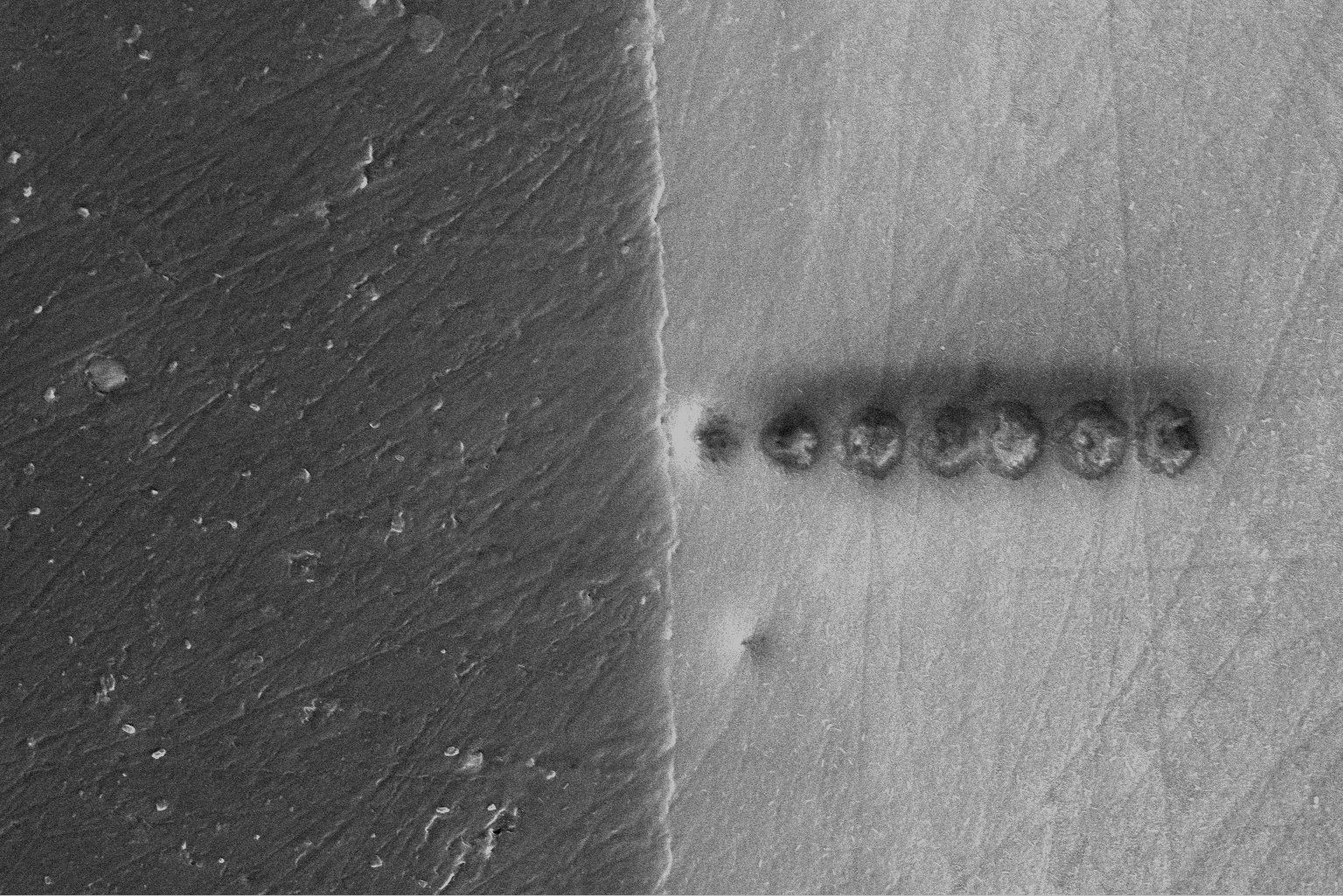
Mansell, Rhodri; Cowburn, Russell; Lesniak, Maciej; Vemulkar, Tarun; Petit, Dorothée CMC; Cheng, Yu; Murphy, Jason
Supporting data for 'Magnetic particles with perpendicular anisotropy for mechanical cancer cell destruction' Journal Article
In: 2017.
@article{mansell2017supporting,

title = {Supporting data for 'Magnetic particles with perpendicular anisotropy for mechanical cancer cell destruction'},

author = {Rhodri Mansell and Russell Cowburn and Maciej Lesniak and Tarun Vemulkar and Dorothée CMC Petit and Yu Cheng and Jason Murphy},

url = {https://www.repository.cam.ac.uk/handle/1810/264210},

year  = {2017},

date = {2017-01-01},

publisher = {University of Cambridge},

abstract = {Supporting data for 'Magnetic particles with perpendicular anisotropy for mechanical cancer cell destruction' published in Scientific Reports. Consisting of magnetic measurement data for figures 1 and 4 and image data for figure 2. Matlab code for generating figure 4b is also included.},

keywords = {},

pubstate = {published},

tppubtype = {article}

}

Robinson, William E; Bassegoda, Arnau; Reisner, Erwin; Hirst, Judy
Oxidation-state-dependent binding properties of the active site in a Mo-containing formate dehydrogenase Journal Article
In: Journal of the American Chemical Society, vol. 139, no. 29, pp. 9927–9936, 2017.
@article{robinson2017oxidation,

title = {Oxidation-state-dependent binding properties of the active site in a Mo-containing formate dehydrogenase},

author = {William E Robinson and Arnau Bassegoda and Erwin Reisner and Judy Hirst},

url = {https://pubs.acs.org/doi/abs/10.1021/jacs.7b03958},

year  = {2017},

date = {2017-01-01},

journal = {Journal of the American Chemical Society},

volume = {139},

number = {29},

pages = {9927--9936},

publisher = {American Chemical Society},

abstract = {Molybdenum-containing formate dehydrogenase H from Escherichia coli (EcFDH-H) is a powerful model system for studies of the reversible reduction of CO2 to formate. However, the mechanism of FDH catalysis is currently under debate, and whether the primary Mo coordination sphere remains saturated or one of the ligands dissociates to allow direct substrate binding during turnover is disputed. Herein, we describe how oxidation-state-dependent changes at the active site alter its inhibitor binding properties. Using protein film electrochemistry, we show that formate oxidation by EcFDH-H is inhibited strongly and competitively by N3–, OCN–, SCN–, NO2–, and NO3–, whereas CO2 reduction is inhibited only weakly and not competitively. During catalysis, the Mo center cycles between the formal Mo(VI)═S and Mo(IV)—SH states, and by modeling chronoamperometry data recorded at different potentials and substrate and inhibitor concentrations, we demonstrate that both formate oxidation and CO2 reduction are inhibited by selective inhibitor binding to the Mo(VI)═S state. The strong dependence of inhibitor-binding affinity on both Mo oxidation state and inhibitor electron-donor strength indicates that inhibitors (and substrates) bind directly to the Mo center. We propose that inhibitors bind to the Mo following dissociation of a selenocysteine ligand to create a vacant coordination site for catalysis and close by considering the implications of our data for the mechanisms of formate oxidation and CO2 reduction.},

keywords = {},

pubstate = {published},

tppubtype = {article}

}

Chen, Kaikai; Juhasz, Matyas; Gularek, Felix; Weinhold, Elmar; Tian, Yu; Keyser, Ulrich F; Bell, Nicholas AW
Ionic Current-Based mapping of short sequence motifs in single DNA molecules using Solid-State nanopores Journal Article
In: Nano letters, vol. 17, no. 9, pp. 5199–5205, 2017.
@article{chen2017ionic,

title = {Ionic Current-Based mapping of short sequence motifs in single DNA molecules using Solid-State nanopores},

author = {Kaikai Chen and Matyas Juhasz and Felix Gularek and Elmar Weinhold and Yu Tian and Ulrich F Keyser and Nicholas AW Bell},

url = {https://pubs.acs.org/doi/abs/10.1021/acs.nanolett.7b01009},

year  = {2017},

date = {2017-01-01},

journal = {Nano letters},

volume = {17},

number = {9},

pages = {5199--5205},

publisher = {American Chemical Society},

abstract = {Nanopore sensors show great potential for rapid, single-molecule determination of DNA sequence information. Here, we develop an ionic current-based method for determining the positions of short sequence motifs in double-stranded DNA molecules with solid-state nanopores. Using the DNA-methyltransferase M.TaqI and a biotinylated S-adenosyl-l-methionine cofactor analogue we create covalently attached biotin labels at 5′-TCGA-3′ sequence motifs. Monovalent streptavidin is then added to bind to the biotinylated sites giving rise to additional current blockade signals when the DNA passes through a conical quartz nanopore. We determine the relationship between translocation time and position along the DNA contour and find a minimum resolvable distance between two labeled sites of ∼200 bp. We then characterize a variety of DNA molecules by determining the positions of bound streptavidin and show that two short genomes can be simultaneously detected in a mixture. Our method provides a simple, generic single-molecule detection platform enabling DNA characterization in an electrical format suited for portable devices for potential diagnostic applications.},

keywords = {},

pubstate = {published},

tppubtype = {article}

}
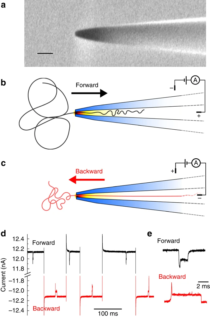
Bell, Nicholas AW; Chen, Kaikai; Ghosal, Sandip; Ricci, Maria; Keyser, Ulrich F
Asymmetric dynamics of DNA entering and exiting a strongly confining nanopore Journal Article
In: Nature communications, vol. 8, no. 1, pp. 1–8, 2017.
@article{bell2017asymmetric,

title = {Asymmetric dynamics of DNA entering and exiting a strongly confining nanopore},

author = {Nicholas AW Bell and Kaikai Chen and Sandip Ghosal and Maria Ricci and Ulrich F Keyser},

url = {https://www.nature.com/articles/s41467-017-00423-9},

year  = {2017},

date = {2017-01-01},

journal = {Nature communications},

volume = {8},

number = {1},

pages = {1--8},

publisher = {Nature Publishing Group},

abstract = {In nanopore sensing, changes in ionic current are used to analyse single molecules in solution. The translocation dynamics of polyelectrolytes is of particular interest given potential applications such as DNA sequencing. In this paper, we determine how the dynamics of voltage driven DNA translocation can be affected by the nanopore geometry and hence the available configurational space for the DNA. Using the inherent geometrical asymmetry of a conically shaped nanopore, we examine how DNA dynamics depends on the directionality of transport. The total translocation time of DNA when exiting the extended conical confinement is significantly larger compared to the configuration where the DNA enters the pore from the open reservoir. By using specially designed DNA molecules with positional markers, we demonstrate that the translocation velocity progressively increases as the DNA exits from confinement. We show that a hydrodynamic model can account for these observations.},

keywords = {},

pubstate = {published},

tppubtype = {article}

}

Krakow, Robert; Bennett, Robbie J; Johnstone, Duncan N; Midgley, Paul A; Hielsher, Ralf; Rae, Catherine MF
Inter-phase Relationships Revealed in 3-Dimensional Orientation Spaces Journal Article
In: Microscopy and Microanalysis, vol. 23, no. S1, pp. 202–203, 2017.
@article{krakow2017inter,

title = {Inter-phase Relationships Revealed in 3-Dimensional Orientation Spaces},

author = {Robert Krakow and Robbie J Bennett and Duncan N Johnstone and Paul A Midgley and Ralf Hielsher and Catherine MF Rae},

url = {https://www.cambridge.org/core/journals/microscopy-and-microanalysis/article/interphase-relationships-revealed-in-3dimensional-orientation-spaces/A4BC2311147429B6DC3782EE09D47C3B},

year  = {2017},

date = {2017-01-01},

journal = {Microscopy and Microanalysis},

volume = {23},

number = {S1},

pages = {202--203},

publisher = {Cambridge University Press},

abstract = {Spatially resolved orientation mapping is increasingly performed using electron microscopy techniques, including: electron backscatter diffraction, transmission Kikuchi diffraction and scanning precession

electron diffraction. The resulting orientation maps contain a wealth of information with crystal phase and orientation specified at each pixel. However, the depth of this data is often underutilized owing to

challenges posed by the analysis of such large quantities of data. In the context of understanding complex and multi-phase materials it is important to characterize inter-phase relationships between

nanoscale precipitates and the surrounding matrix, which both affect properties and are indicative of formation pathways. Revealing inter-phase relationships requires statistical assessment of the orientation relationship across the phase boundary, the spatial occurrence of particular boundaries and the surfaces of contact at interfaces. Analysis procedures that highlight relationships in both spatial and orientation dimensions are therefore required. Here, we present an approach to revealing inter-phase relationships, based on considering orientation data in 3-dimensional vector spaces constrained to fundamental zones defined by the crystal symmetry of both crystals.},

keywords = {},

pubstate = {published},

tppubtype = {article}

}
electron diffraction. The resulting orientation maps contain a wealth of information with crystal phase and orientation specified at each pixel. However, the depth of this data is often underutilized owing to
challenges posed by the analysis of such large quantities of data. In the context of understanding complex and multi-phase materials it is important to characterize inter-phase relationships between
nanoscale precipitates and the surrounding matrix, which both affect properties and are indicative of formation pathways. Revealing inter-phase relationships requires statistical assessment of the orientation relationship across the phase boundary, the spatial occurrence of particular boundaries and the surfaces of contact at interfaces. Analysis procedures that highlight relationships in both spatial and orientation dimensions are therefore required. Here, we present an approach to revealing inter-phase relationships, based on considering orientation data in 3-dimensional vector spaces constrained to fundamental zones defined by the crystal symmetry of both crystals.
Sunde, Jonas K; Johnstone, Duncan N; Marioara, Calin D; van Helvoort, Antonius TJ; Midgley, Paul A; Holmestad, Randi
Scanning Precession Electron Diffraction Study of Hybrid Precipitates in a 6xxx Series Aluminium Alloy Journal Article
In: Microscopy and Microanalysis, vol. 23, no. S1, pp. 114–115, 2017.
@article{sunde2017scanning,

title = {Scanning Precession Electron Diffraction Study of Hybrid Precipitates in a 6xxx Series Aluminium Alloy},

author = {Jonas K Sunde and Duncan N Johnstone and Calin D Marioara and Antonius TJ van Helvoort and Paul A Midgley and Randi Holmestad},

url = {https://www.cambridge.org/core/journals/microscopy-and-microanalysis/article/scanning-precession-electron-diffraction-study-of-hybrid-precipitates-in-a-6xxx-series-aluminium-alloy/0ACABFE3E679044756F4985CF4C9A229},

year  = {2017},

date = {2017-01-01},

journal = {Microscopy and Microanalysis},

volume = {23},

number = {S1},

pages = {114--115},

publisher = {Cambridge University Press},

abstract = {Obtaining reliable and statistical assessment of nanoscale precipitates to link the microstructure with material properties is a primary objective of transmission electron microscopy (TEM) studies of agehardenable aluminium (Al) alloys. Here, it is demonstrated that nanometer-resolution scanning precession electron diffraction (SPED) [1] combined with versatile post-facto computational analysis

provides a route to extract improved precipitate statistics in Al alloys. The approach developed is automated, and offers more objective assessment compared to conventional imaging procedures which

require manual identification, counting and measurements. The material studied here is a common 6xxx series industrial alloy (6082, Al-0.72Mg-0.88Si-0.03Cu (wt %)) and the methods developed are generic

and applicable to any material containing nanoscale precipitates.},

keywords = {},

pubstate = {published},

tppubtype = {article}

}
provides a route to extract improved precipitate statistics in Al alloys. The approach developed is automated, and offers more objective assessment compared to conventional imaging procedures which
require manual identification, counting and measurements. The material studied here is a common 6xxx series industrial alloy (6082, Al-0.72Mg-0.88Si-0.03Cu (wt %)) and the methods developed are generic
and applicable to any material containing nanoscale precipitates.
Johnstone, Duncan N; Midgley, Paul A
The Microstructure of Pharmaceutical Materials Revealed by Scanning Electron Diffraction Journal Article
In: Microscopy and Microanalysis, vol. 23, no. S1, pp. 1192–1193, 2017.
@article{johnstone2017microstructure,

title = {The Microstructure of Pharmaceutical Materials Revealed by Scanning Electron Diffraction},

author = {Duncan N Johnstone and Paul A Midgley},

url = {https://www.cambridge.org/core/journals/microscopy-and-microanalysis/article/microstructure-of-pharmaceutical-materials-revealed-by-scanning-electron-diffraction/AC52A1F2F736891CC40A66DF405521FE},

year  = {2017},

date = {2017-01-01},

journal = {Microscopy and Microanalysis},

volume = {23},

number = {S1},

pages = {1192--1193},

publisher = {Cambridge University Press},

abstract = {The microstructure of pharmaceutical materials influences important physical properties [1] such as compaction behavior, dissolution rate, bioavailability, hygroscopicity, dehydration, chemical stability

and physical stability. However, few techniques are capable of directly revealing the underlying microstructure and associated defects. Transmission electron microscopy (TEM) provides an obvious

means to image and characterize microstructure and defects. Whilst previous TEM studies of pharmaceutical materials have been insightful [2] the level of detail attained has been necessarily limited

because of the sensitivity of the sample to damage by electron irradiation. Scanning electron diffraction (SED) provides a new paradigm in which the data required for detailed analysis is acquired rapidly and

before the specimen is destroyed. Insight is gleaned post-facto through the application of versatile computational analysis enabling maximal information to be extracted from the data.},

keywords = {},

pubstate = {published},

tppubtype = {article}

}
and physical stability. However, few techniques are capable of directly revealing the underlying microstructure and associated defects. Transmission electron microscopy (TEM) provides an obvious
means to image and characterize microstructure and defects. Whilst previous TEM studies of pharmaceutical materials have been insightful [2] the level of detail attained has been necessarily limited
because of the sensitivity of the sample to damage by electron irradiation. Scanning electron diffraction (SED) provides a new paradigm in which the data required for detailed analysis is acquired rapidly and
before the specimen is destroyed. Insight is gleaned post-facto through the application of versatile computational analysis enabling maximal information to be extracted from the data.
Johnstone, Duncan N; van Helvoort, Antonius TJ; Midgley, Paul A
Nanoscale strain tomography by scanning precession electron diffraction Journal Article
In: Microscopy and Microanalysis, vol. 23, no. S1, pp. 1710–1711, 2017.
@article{johnstone2017nanoscale,

title = {Nanoscale strain tomography by scanning precession electron diffraction},

author = {Duncan N Johnstone and Antonius TJ van Helvoort and Paul A Midgley},

url = {https://www.cambridge.org/core/journals/microscopy-and-microanalysis/article/nanoscale-strain-tomography-by-scanning-precession-electron-diffraction/A300A71406DFDFD793D889F07D9DF472#metrics},

year  = {2017},

date = {2017-01-01},

journal = {Microscopy and Microanalysis},

volume = {23},

number = {S1},

pages = {1710--1711},

publisher = {Cambridge University Press},

abstract = {Strain within materials can be engineered on the nanoscale to control physical properties from electronics to mechanics. Methods for mapping this strain experimentally include both X-ray diffraction

and electron microscopy techniques. X-ray techniques such as Laue micro-diffraction [1] and coherent X-ray diffraction [2] offer spatial resolution of ~200 nm and ~30 nm respectively and electron backscatter diffraction [3] reduces this only slightly. Spatial resolution down to ~1 nm is obtained only via (scanning) transmission electron microscopy ((S)TEM) methods [4] such as geometric phase analysis, dark-field holography and scanning (precession) electron diffraction (SPED). These (S)TEM techniques have been evaluated extensively for strain mapping in semiconductor devices [4] and it has emerged that SPED offers several benefits in terms of precision, ease of implementation and balance between spatial resolution and field of view. Further, the SPED approach is highly versatile and can be applied to a wide range of materials systems and geometries.

},

keywords = {},

pubstate = {published},

tppubtype = {article}

}
and electron microscopy techniques. X-ray techniques such as Laue micro-diffraction [1] and coherent X-ray diffraction [2] offer spatial resolution of ~200 nm and ~30 nm respectively and electron backscatter diffraction [3] reduces this only slightly. Spatial resolution down to ~1 nm is obtained only via (scanning) transmission electron microscopy ((S)TEM) methods [4] such as geometric phase analysis, dark-field holography and scanning (precession) electron diffraction (SPED). These (S)TEM techniques have been evaluated extensively for strain mapping in semiconductor devices [4] and it has emerged that SPED offers several benefits in terms of precision, ease of implementation and balance between spatial resolution and field of view. Further, the SPED approach is highly versatile and can be applied to a wide range of materials systems and geometries.

Dehmel, Raphael; Dolan, James A; Gu, Yibei; Wiesner, Ulrich; Wilkinson, Timothy D; Baumberg, Jeremy J; Steiner, Ullrich; Wilts, Bodo D; Gunkel, Ilja
Optical imaging of large gyroid grains in block copolymer templates by confined crystallization Journal Article
In: Macromolecules, vol. 50, no. 16, pp. 6255–6262, 2017.
@article{dehmel2017optical,

title = {Optical imaging of large gyroid grains in block copolymer templates by confined crystallization},

author = {Raphael Dehmel and James A Dolan and Yibei Gu and Ulrich Wiesner and Timothy D Wilkinson and Jeremy J Baumberg and Ullrich Steiner and Bodo D Wilts and Ilja Gunkel},

url = {https://pubs.acs.org/doi/abs/10.1021/acs.macromol.7b01528},

year  = {2017},

date = {2017-01-01},

journal = {Macromolecules},

volume = {50},

number = {16},

pages = {6255--6262},

publisher = {American Chemical Society},

abstract = {Block copolymer (BCP) self-assembly is a promising route to manufacture functional nanomaterials for applications from nanolithography to optical metamaterials. Self-assembled cubic morphologies cannot, however, be conveniently optically characterized in the lab due to their structural isotropy. Here, the aligned crystallization behavior of a semicrystalline-amorphous polyisoprene-b-polystyrene-b-poly(ethylene oxide) (ISO) triblock terpolymer was utilized to visualize the grain structure of the cubic microphase-separated morphology. Upon quenching from a solvent swollen state, ISO first self-assembles into an alternating gyroid morphology, in the confinement of which the PEO crystallizes preferentially along the least tortuous pathways of the single gyroid morphology with grain sizes of hundreds of micrometers. Strikingly, the resulting anisotropic alignment of PEO crystallites gives rise to a unique optical birefringence of the alternating gyroid domains, which allows imaging of the self-assembled grain structure by optical microscopy alone. This study provides insight into polymer crystallization within a tortuous three-dimensional network and establishes a useful method for the optical visualization of cubic BCP morphologies that serve as functional nanomaterial templates.},

keywords = {},

pubstate = {published},

tppubtype = {article}

}

Jussila, Henri; Albrow-Owen, Tom; Yang, He; Hu, Guohua; Aksimsek, Sinan; Granqvist, Niko; Lipsanen, Harri; Howe, Richard CT; Sun, Zhipei; Hasan, Tawfique
New approach for thickness determination of solution-deposited graphene thin films Journal Article
In: ACS omega, vol. 2, no. 6, pp. 2630–2638, 2017.
@article{jussila2017new,

title = {New approach for thickness determination of solution-deposited graphene thin films},

author = {Henri Jussila and Tom Albrow-Owen and He Yang and Guohua Hu and Sinan Aksimsek and Niko Granqvist and Harri Lipsanen and Richard CT Howe and Zhipei Sun and Tawfique Hasan},

url = {https://pubs.acs.org/doi/abs/10.1021/acsomega.7b00336},

year  = {2017},

date = {2017-01-01},

journal = {ACS omega},

volume = {2},

number = {6},

pages = {2630--2638},

publisher = {American Chemical Society},

abstract = {Solution processing-based fabrication techniques such as liquid phase exfoliation may enable economically feasible utilization of graphene and related nanomaterials in real-world devices in the near future. However, measurement of the thickness of the thin film structures fabricated by these approaches remains a significant challenge. By using surface plasmon resonance (SPR), a simple, accurate, and quick measurement of the deposited thickness for inkjet-printed graphene thin films is reported here. We show that the SPR technique is convenient and well-suited for the measurement of thin films formulated from nanomaterial inks, even at sub-10 nm thickness. We also demonstrate that the analysis required to obtain results from the SPR measurements is significantly reduced compared to that required for atomic force microscopy (AFM) or stylus profilometer, and much less open to interpretation. The gathered data implies that the film thickness increases linearly with increasing number of printing repetitions. In addition, SPR also reveals the complex refractive index of the printed thin films composed of exfoliated graphene flakes, providing a more rigorous explanation of the optical absorption than that provided by a combination of AFM/profilometer and the extinction coefficient of mechanically exfoliated graphene flakes. Our results suggest that the SPR method may provide a new pathway for the thickness measurement of thin films fabricated from any nanomaterial containing inks.},

keywords = {},

pubstate = {published},

tppubtype = {article}

}

Liu, Ke; Lihter, Martina; Sarathy, Aditya; Caneva, Sabina; Qiu, Hu; Deiana, Davide; Tileli, Vasiliki; Alexander, Duncan TL; Hofmann, Stephan; Dumcenco, Dumitru; others,
Geometrical effect in 2D nanopores Journal Article
In: Nano letters, vol. 17, no. 7, pp. 4223–4230, 2017.
@article{liu2017geometrical,

title = {Geometrical effect in 2D nanopores},

author = {Ke Liu and Martina Lihter and Aditya Sarathy and Sabina Caneva and Hu Qiu and Davide Deiana and Vasiliki Tileli and Duncan TL Alexander and Stephan Hofmann and Dumitru Dumcenco and others},

url = {https://pubs.acs.org/doi/abs/10.1021/acs.nanolett.7b01091},

year  = {2017},

date = {2017-01-01},

journal = {Nano letters},

volume = {17},

number = {7},

pages = {4223--4230},

publisher = {American Chemical Society},

abstract = {A long-standing problem in the application of solid-state nanopores is the lack of the precise control over the geometry of artificially formed pores compared to the well-defined geometry in their biological counterpart, that is, protein nanopores. To date, experimentally investigated solid-state nanopores have been shown to adopt an approximately circular shape. In this Letter, we investigate the geometrical effect of the nanopore shape on ionic blockage induced by DNA translocation using triangular h-BN nanopores and approximately circular molybdenum disulfide (MoS2) nanopores. We observe a striking geometry-dependent ion scattering effect, which is further corroborated by a modified ionic blockage model. The well-acknowledged ionic blockage model is derived from uniform ion permeability through the 2D nanopore plane and hemisphere like access region in the nanopore vicinity. On the basis of our experimental results, we propose a modified ionic blockage model, which is highly related to the ionic profile caused by geometrical variations. Our findings shed light on the rational design of 2D nanopores and should be applicable to arbitrary nanopore shapes.},

keywords = {},

pubstate = {published},

tppubtype = {article}

}
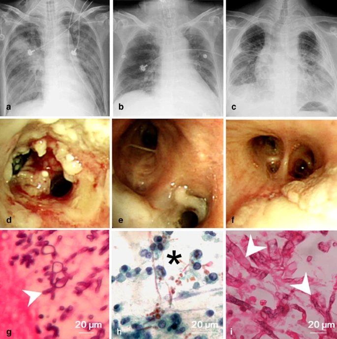
Lin, Chun-Yu; Liu, Wei-Lun; Chang, Che-Chia; Chang, Hou-Tai; Hu, Han-Chung; Kao, Kuo-chin; Chen, Ning-Hung; Chen, Ying-Jen; Yang, Cheng-Ta; Huang, Chung-Chi; others,
Invasive fungal tracheobronchitis in mechanically ventilated critically ill patients: underlying conditions, diagnosis, and outcomes Journal Article
In: Annals of intensive care, vol. 7, no. 1, pp. 1–7, 2017.
@article{lin2017invasive,

title = {Invasive fungal tracheobronchitis in mechanically ventilated critically ill patients: underlying conditions, diagnosis, and outcomes},

author = {Chun-Yu Lin and Wei-Lun Liu and Che-Chia Chang and Hou-Tai Chang and Han-Chung Hu and Kuo-chin Kao and Ning-Hung Chen and Ying-Jen Chen and Cheng-Ta Yang and Chung-Chi Huang and others},

url = {https://link.springer.com/article/10.1186/s13613-016-0230-9},

year  = {2017},

date = {2017-01-01},

journal = {Annals of intensive care},

volume = {7},

number = {1},

pages = {1--7},

publisher = {SpringerOpen},

abstract = {This study included 31 patients who had been diagnosed as having IFT, comprising 24 men and 7 women with a mean age of 64.7 ± 13.7 years. All patients developed respiratory failure and received mechanical ventilation before diagnosis. Eighteen (58.1%) patients had diabetes mellitus, and 12 (38.7%) had chronic lung disease. Four (12.9%) patients had hematologic disease, and none of the patients had neutropenia. Twenty-five (80.6%) patients were diagnosed as having proven IFT, and the remaining patients had probable IFT. Aspergillus spp. (61.3%) were the most common pathogenic species, followed by Mucorales (25.8%) and Candida spp. (6.5%). The diagnoses in six (19.4%) patients were confirmed only through bronchial biopsy and histopathological examination, whereas their cultures of bronchoalveolar lavage fluid were negative for fungi. The overall in-hospital mortality rate was 93.5%.},

keywords = {},

pubstate = {published},

tppubtype = {article}

}
Weber, Isabell P; Rana, Mrinal; Thomas, Peter BM; Dimov, Ivan B; Franze, Kristian; Rajan, Madhavan S
Effect of vital dyes on human corneal endothelium and elasticity of Descemet’s membrane Journal Article
In: PloS one, vol. 12, no. 9, pp. e0184375, 2017.
@article{weber2017effect,

title = {Effect of vital dyes on human corneal endothelium and elasticity of Descemet’s membrane},

author = {Isabell P Weber and Mrinal Rana and Peter BM Thomas and Ivan B Dimov and Kristian Franze and Madhavan S Rajan},

url = {https://journals.plos.org/plosone/article?id=10.1371/journal.pone.0184375},

year  = {2017},

date = {2017-01-01},

journal = {PloS one},

volume = {12},

number = {9},

pages = {e0184375},

publisher = {Public Library of Science San Francisco, CA USA},

abstract = {The purpose of this study was to evaluate the effects of vital dyes on human Descemet's membranes (DMs) and endothelia. DMs of 25 human cadaveric corneas with research consent were treated with dyes routinely used in Descemet membrane endothelial keratoplasty (DMEK), 0.05% Trypan blue (TB) or a combination of 0.15% Trypan blue, 0.025% Brilliant blue and 4% Polyethylene glycol (commercial name Membrane Blue Dual; MB). The effects of these two dyes on (i) endothelial cell viability, (ii) DM mechanical properties as assessed by atomic force microscopy, and iii) qualitative DM dye retention were tested for two varying exposure times (one or four minutes). No significant differences in cell toxicity were observed between treatments with TB and MB at the two different exposure times (P = 0.21). Further, both dyes led to a significant increase in DM stiffness: exposure to TB and MB for one minute increased the apparent elastic modulus of the DM by 11.2% (P = 8*10−3) and 17.7%, respectively (P = 4*10−6). A four-minute exposure led to an increase of 8.6% for TB (P = 0.004) and 13.6% for MB (P = 0.03). Finally, at 25 minutes, the dye retention of the DM was considerably better for MB compared to TB. Taken together, a one-minute exposure to MB was found to improve DM visibility compared to TB, with a significant increase in DM stiffness and without detrimental effects on endothelial cell viability. The use of MB could therefore improve (i) visibility of the DM scroll, and (ii) intraoperative unfolding, enhancing the probability of successful DMEK surgery.},

keywords = {},

pubstate = {published},

tppubtype = {article}

}
Choi, Yeon Sik; Jing, Qingshen; Datta, Anuja; Boughey, Chess; Kar-Narayan, Sohini
A triboelectric generator based on self-poled Nylon-11 nanowires fabricated by gas-flow assisted template wetting Journal Article
In: Energy & Environmental Science, vol. 10, no. 10, pp. 2180–2189, 2017.
@article{choi2017triboelectric,

title = {A triboelectric generator based on self-poled Nylon-11 nanowires fabricated by gas-flow assisted template wetting},

author = {Yeon Sik Choi and Qingshen Jing and Anuja Datta and Chess Boughey and Sohini Kar-Narayan},

url = {https://pubs.rsc.org/no/content/articlehtml/2017/ee/c7ee01292f},

year  = {2017},

date = {2017-01-01},

journal = {Energy & Environmental Science},

volume = {10},

number = {10},

pages = {2180--2189},

publisher = {Royal Society of Chemistry},

abstract = {Triboelectric generators have emerged as potential candidates for mechanical energy harvesting, relying on motion-generated surface charge transfer between materials with different electron affinities. In this regard, synthetic organic materials with strong electron-donating tendencies are far less common than their electron-accepting counterparts. Nylons are notable exceptions, with odd-numbered Nylons such as Nylon-11, exhibiting electric polarisation that could further enhance the surface charge density crucial to triboelectric generator performance. However, the fabrication of Nylon-11 in the required polarised δ′-phase typically requires extremely rapid crystallisation, such as melt-quenching, as well as “poling” via mechanical stretching and/or large electric fields for dipolar alignment. Here, we propose an alternative one-step, near room-temperature fabrication method, namely gas-flow assisted nano-template (GANT) infiltration, by which highly crystalline “self-poled” δ′-phase Nylon-11 nanowires are grown from solution within nanoporous anodised aluminium oxide (AAO) templates. Our gas-flow assisted method allows for controlled crystallisation of the δ′-phase of Nylon-11 through rapid solvent evaporation and an artificially generated extreme temperature gradient within the nanopores of the AAO template, as accurately predicted by finite-element simulations. Furthermore, preferential crystal orientation originating from template-induced nano-confinement effects leads to self-poled δ′-phase Nylon-11 nanowires with higher surface charge distribution than melt-quenched Nylon-11 films, as observed by Kelvin probe force microscopy (KPFM). Correspondingly, a triboelectric nanogenerator (TENG) device based on as-grown templated Nylon-11 nanowires fabricated via GANT infiltration showed a ten-fold increase in output power density as compared to an aluminium-based triboelectric generator, when subjected to identical mechanical excitations.},

keywords = {},

pubstate = {published},

tppubtype = {article}

}

Griffiths, Matthew; Niblett, Samuel P; Wales, David J
Optimal alignment of structures for finite and periodic systems Journal Article
In: Journal of chemical theory and computation, vol. 13, no. 10, pp. 4914–4931, 2017.
@article{griffiths2017optimal,

title = {Optimal alignment of structures for finite and periodic systems},

author = {Matthew Griffiths and Samuel P Niblett and David J Wales},

url = {https://pubs.acs.org/doi/abs/10.1021/acs.jctc.7b00543},

year  = {2017},

date = {2017-01-01},

journal = {Journal of chemical theory and computation},

volume = {13},

number = {10},

pages = {4914--4931},

publisher = {American Chemical Society},

abstract = {Finding the optimal alignment between two structures is important for identifying the minimum root-mean-square distance (RMSD) between them and as a starting point for calculating pathways. Most current algorithms for aligning structures are stochastic, scale exponentially with the size of structure, and the performance can be unreliable. We present two complementary methods for aligning structures corresponding to isolated clusters of atoms and to condensed matter described by a periodic cubic supercell. The first method (Go-PERMDIST), a branch and bound algorithm, locates the global minimum RMSD deterministically in polynomial time. The run time increases for larger RMSDs. The second method (FASTOVERLAP) is a heuristic algorithm that aligns structures by finding the global maximum kernel correlation between them using fast Fourier transforms (FFTs) and fast SO(3) transforms (SOFTs). For periodic systems, FASTOVERLAP scales with the square of the number of identical atoms in the system, reliably finds the best alignment between structures that are not too distant, and shows significantly better performance than existing algorithms. The expected run time for Go-PERMDIST is longer than FASTOVERLAP for periodic systems. For finite clusters, the FASTOVERLAP algorithm is competitive with existing algorithms. The expected run time for Go-PERMDIST to find the global RMSD between two structures deterministically is generally longer than for existing stochastic algorithms. However, with an earlier exit condition, Go-PERMDIST exhibits similar or better performance.},

keywords = {},

pubstate = {published},

tppubtype = {article}

}